Fetal Neurosonogram: Detailed Ultrasound of the Baby's Brain and Spine
A fetal neurosonogram is a specialized ultrasound examination designed to evaluate the development of the baby's brain and spinal cord during pregnancy. This scan provides detailed images of the central nervous system and helps doctors detect possible abnormalities at an early stage.
This advanced ultrasound is often recommended when routine pregnancy scans suggest possible concerns related to the brain or spine. It may also be advised for pregnancies that have certain risk factors, such as maternal infections, genetic conditions, or a family history of neurological disorders.
By closely examining the fetal nervous system, a neurosonogram helps doctors confirm whether the baby's brain structures are developing normally and allows them to plan appropriate monitoring or care if required.
What Does a Fetal Neurosonogram Involve?
Detailed Brain Examination: The sonographer carefully evaluates different parts of the fetal brain, including the ventricles, cerebellum, and other important structures.
Spinal Cord Assessment: The spine is examined to ensure proper alignment and development of the spinal cord.
Measurements: Specific measurements are taken to assess the size, shape, and development of the brain structures.
Detection of Abnormalities: The scan may help identify conditions such as hydrocephalus, spina bifida, brain malformations, or other neurological abnormalities.
When is a Fetal Neurosonogram Recommended?
Family History of Neurological Disorders: If there is a known history of neurological conditions in the family.
Abnormal Findings on Routine Ultrasound: If earlier scans show possible concerns related to the brain or spine.
Maternal Health Conditions: Certain maternal conditions, such as infections during pregnancy or uncontrolled diabetes, may increase the risk of fetal neurological problems.
Specific Clinical Concerns: Doctors may recommend this scan if there are particular concerns about the baby's neurological development.
What to Expect During the Procedure
Preparation: Usually no special preparation is required before the scan.
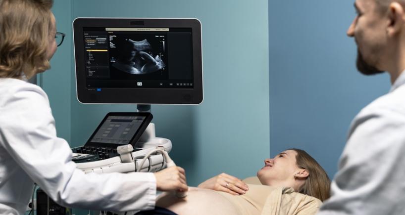
The Procedure: A small amount of gel is applied to the abdomen and a handheld ultrasound probe is used to capture detailed images of the fetal brain and spine.
Duration: The examination typically takes about 30 to 45 minutes, depending on the baby's position and the level of detail required.
Important: While fetal neurosonography provides detailed information about the developing nervous system, it may not detect every possible neurological condition. Your healthcare provider will discuss the findings and advise you about any further evaluation if needed.