Fetal Echocardiography: Detailed Evaluation of the Baby’s Heart
Fetal echocardiography is a specialized ultrasound test used to examine the heart of a developing baby during pregnancy. This scan focuses specifically on the structure and function of the fetal heart, helping doctors evaluate how the heart chambers, valves, and blood vessels are forming and working.
The test provides detailed images of the baby’s heart and helps detect possible congenital heart defects or abnormalities. Early diagnosis of heart conditions allows doctors to plan appropriate monitoring, treatment, and delivery care if necessary.
Fetal echocardiography is usually performed by specialists using advanced ultrasound technology to obtain a detailed assessment of the baby’s cardiovascular system.
When is Fetal Echocardiography Recommended?
Family History of Heart Disease: If there is a family history of congenital heart defects or other cardiac conditions.
Maternal Health Conditions: Certain maternal conditions, such as diabetes, autoimmune disorders, or infections during pregnancy, may increase the risk of fetal heart abnormalities.
Abnormal Ultrasound Findings: If routine pregnancy ultrasounds suggest possible concerns related to the baby’s heart or circulation.
High-Risk Pregnancies: Doctors may recommend fetal echocardiography in pregnancies considered high risk or when there are specific concerns about heart development.
What Happens During a Fetal Echocardiography Scan?
Detailed Heart Examination: The sonographer carefully evaluates the four chambers of the fetal heart, the heart valves, and the major blood vessels.
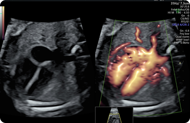
Blood Flow Assessment: Doppler ultrasound may be used to observe the direction and speed of blood flow within the heart.
Heart Measurements: The size, rhythm, and overall function of the heart are assessed to ensure normal development.
Detection of Abnormalities: The scan can help identify certain heart defects, rhythm disturbances (arrhythmias), or structural abnormalities.
What to Expect During the Procedure
Preparation: Usually no special preparation is required before the scan.
The Procedure: A small amount of gel is applied to the abdomen, and a handheld ultrasound probe is used to obtain images of the baby's heart.
Duration: The examination generally takes around 30 to 45 minutes, depending on the baby’s position and the level of detail required.
Important: While fetal echocardiography is highly useful for evaluating the baby's heart, it may not detect every possible heart condition. Your doctor will explain the findings and guide you if further monitoring or evaluation is required.